


|
|
Northeast Branch Newsletter
|
|
|
Number 127
|
December 2005
|
|
|
Membership News
Dues reminders were mailed in November 2005. Please make the necessary corrections and return to the Treasurer. The mailing labels on announcements, etc. will reflect all updates. We have created a membership database in order to send items of interest more easily and rapidly by E-mail, therefore don't forget to include your E-mail address. Please notify Irene H. George, Secretary, of any changes at (617) 983-6371.
Membership in the national branch automatically makes you a member of the local branch in some organizations, but this is NOT the case in the ASM. You may be both a National Member and a NEB member, but you have to join each individually. The Northeast Branch membership currently has 250 paid members, which includes: 21 Emeritus, 4 Honorary and 9 Student members.
back to top
Council Meeting Schedule, 2006
Council Meetings this year will be held at the State Laboratory Institute in Jamaica Plain. Members and all interested microbiologists and scientists are welcome to attend. Please notify Irene H. George at (617) 983-6371 in advance. The next 2006 Council Meetings are scheduled for January 10, March 1, May 3 and June (date to be announced).
back to top
Visit Our Web Site!!
The NEB has established a home page on the World Wide Web where all current events and the Newsletter are available. ASM has also established a Branch Meetings page. Visit us via the ASM Home Page or directly at: http://www.asm.org/Branch/brNoE/index.shtml.
back to top
Council Elections
Congratulations to the following newly elected officers: President-Elect, Jeffrey Klinger; Secretary, Irene H. George; National Councilor, Paulette Howarth; Alternate National Councilor, Frank Scarano; and Local Councilor, Sandra Smole. We look forward to working with you in the coming year!
back to top
Joining a Branch
Have you taken advantage of everything your local ASM Branch has to offer? ASM's thirty-five Branches are organized geographically. Each Branch is individually chartered, and elects their own officers, sets their own rules, and determines their own membership criteria. Membership in the National ASM is not required to join a Branch, but National membership is encouraged. Branches offer members an opportunity to network, search for a job, and become involved in science in the community. Branches also hold meetings with ASM Foundation Lecturers, offer opportunities for students to present papers, organize collegial dinners, and foster science at the local level. If you are a National ASM member, you can join a local Branch through the National Branch when you renew your membership this year, or you can join online at http://eStore.asm.org. Beginning in September 2005, National started to collect Branch Full Member dues in conjunction with the processing of 2006 ASM ONLINE Full Member Applications. Members can thus add the amount of the dues for that Branch to their payment to National. On a quarterly basis, Membership Services will send Branches checks in the amounts owed for Branch dues paid through National. Names and contact information for people joining Branches will be sent to the Branches every month. The ASM operates on a calendar year basis, January through December, and collects dues beginning the previous September. You can also join by contacting your local Branch representative (irene.george@state.ma.us).
back to top
Science Fair Winners
The NEB annually donates an award of $100 to each of five MA regional fairs and the VT science fair, while the MA Science Fair receives $200. Following are this year's winners of the NEB awards and their projects. Congratulations again for your outstanding work. We would like to thank Council members Gregory Reppucci, Paulette Howarth, and other NEB members for volunteering to judge at these fairs. We were unable to obtain the names of winners at the Region 4 Somerville Science Fair and VT State Science and Mathematics Fair at this time. Recipients of this year's science fair awards were:
2005 Massachusetts State Science Fair: Jason Hu, Boston Latin Academy, with Genomic Signatures of Prokaryotes by Chaos Game Representation.
Region 2: Worcester Regional Science and Engineering Fair: E. coli Reduction on Cutting Boards, Kristen Baldiga, Hopkinton High School.
Region 3: Bristol Community College-Rensselaer Polytechnic Institute Regional Science Fair, RNA: Mediated Genetic Analysis of Epstein-Barr Virus Lymphoblastoid Cell Lines, Shahrir A. Khan, 12th grade, Attleboro High School.
Region 5: The South Shore Regional Science Fair had two winners who took second place: Neil T. Forrester, grade 11, Falmouth High School with Chemotaxis in E. coli and Daniel Green, grade 11, Upper Cape Regional Technical School, Bourne, with Ghost Crypt.
Region 6: Boston Regional Science Fair: the winner was Lily Silayeva, grade 12, Boston Latin School, with Effects of Dystroglycan Deficiency on Other Proteins in a Cell.
back to top
"Microbe" Coming in January
As announced in October, ASM News magazine will be called Microbe beginning in January 2006. It will remain the news magazine of ASM, and will continue to be a member benefit. The content and format will remain the same. The print version of the magazine will be sent to all U.S. members as a part of the member ship fee and to all international members who pay the shipping charge of $10. The goal of changing the name is to raise the profile of the magazine by making it clear that it contains more than just ASM-related material.
The new Web address of the magazine will be http://www.asm.org/microbe/, and access to most sections will be open to both members and nonmembers beginning in January 2006.
.
Michael Goldberg
Editor in Chief, Microbe
back to top
Programs in Review-2005
The New Chief of Staph and Others That Want To Be
The New Chief of Staph and Others that Want to Be: An Overview of Antibiotic Resistance was presented on December 12, 2005 by Stephen Brecher, PhD, Director of Microbiology, VA Boston Healthcare System, W. Roxbury, MA. An audience of eighty-two people attended the meeting held at Vinny Testa's in Dedham, MA.
Dr. Brecher began with the history of penicillin, when in 1928, Sir Alexander Fleming was studying staphylococcal lysis and observed a mold inhibiting S. aureus on some of his glass Petri dishes. Fleming worked on this observation from 1928-1938, but never made enough of the inhibitory molecule. Sulfa drugs with which bacterial infections were treated came along in 1935. The British returned to the project of penicillin production when war clouds appeared over Europe, turning the penicillin project over to Howard Florey and Ernest Chain at Oxford University. After considerable effort, they were able to produce enough pure penicillin to treat mice infected with S. aureus. In 1941 they treated their first patient, constable Albert Alexander, who started to recover, but died when they ran out of drug. Meanwhile, in 1940-41 the war was raging in Europe, and the British sent Florey to the US to convince our government to back large-scale penicillin production. There was some interest and a few companies started to produce it, but it was not a high priority project.
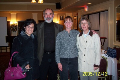
(L to R) Paula Aronson, Stephen Brecher, PhD,
Dianne Fitzsimmons and Elizabeth Leonard,
VA Medical Center, W., Roxbury, MA
Penicillin was made famous by the Coconut Grove Night Club fire in the South End of Boston in 1942. Holy Cross had upset Boston College and the club was filled to capacity; the fire killed 492 people and many were severely burned. At that time sulfa was used for infections and plasma for dehydration and shock, however, patients getting sulfa were now getting infections not controlled by sulfa. A call, with government permission, went to Merck in Rahway, NJ, who rushed 32 liters of fluid medium in which Penicillium had been grown and the people who were treated with this recovered. This "miracle drug" was publicized for several months by the radio and press to the point where the US government realized it was valuable, and instructed the pharmaceutical industry to work on it. Before the fire, only about one hundred Americans had received penicillin. Urine was collected from these people and the drug recrystallized and reused. At that time one dose consisted of 10,000 units. Americans improved the method of production to the point that by D-Day, there was enough penicillin for every wounded American who needed it. Fleming, in 1945, also worked with S. aureus mutants that grew in increasing concentrations of penicillin. In 1946, when an oral over-the-counter form of the drug was available, Fleming wrote of the danger of self-medication, noting that taking small doses may cause resistance.
Dr. Brecher reminded us that bacteria are the dominant species on earth, with a rapid multiplication and a natural mutation rate. They easily transfer or move genes, allowing them to survive, change and flourish under pressure. They obviously will keep and transfer genes that are advantageous to the species, such as resistance genes that arise by mutation and are transferred by plasmids on transposons. The human body has 10 13 human cells and 10 14 bacteria cells. If an infection or antibiotics wipe out normal bacterial flora, C. difficile, Candida and other bacteria, previously held in check will flourish.
The old Chief of Staph was S. aureus said Dr. Brecher. We now have so many mutations that we now use initials for them instead of names, such as MRSA, VISA, GISA, VRSA, and CA-MRSA. This is bacterial evolution and the bugs are fighting back! And we have been using antibiotics for only sixty-three years!
Soon after the introduction of penicillin, the first penicillin resistant strains of S. aureus appeared. In the 50's there were reports of 50% of S. aureus being resistant to penicillin. Semi-synthetic penicillinase-resistant penicillins were produced in 1959, but after only two years of use, the first methicillin resistant MRSA strain appeared (1961). Now 30-70% of all S. aureus are methicillin resistant (MRSA) and are difficult to treat. Vancomycin, produced in the 50's, is considered one of the last lines of defense against serious gram positive infections. Infrequently used in the past, there were no organisms resistant to it, but due to its current widespread use, we have vancomycin intermediate (VISA) strains. These were first reported in 1995 and eight cases were confirmed in the US in 2002. In England in 1991 trans-conjugation, using the vanA gene from S. fecalis, was used to make a S. aureus strain highly resistant to vancomycin (MRSA), and the first fully resistant S. aureus to naturally acquire vanA from enterococci was reported in MMWR in July, 2002. This patient had both vancomycin resistant enterococci (VRE) and VRSA; the MIC was >1024. There have been only five cases of VRSA in about three years to date. Infections with enterococci, which are intrinsically resistant to antibiotics, are also treated with vancomycin. However, (VRE) were reported in England in 1988, and the United States in 1989. The vanA gene that confers high level resistance is seen mostly in S. fecalis and M. faecium; but S. fecalis passes it more readily.
"So who is the new Chief of Staph?" asked Dr. Brecher. This would be community acquired MRSA (CA-MRSA)! Until a few years ago, MRSA meant hospital acquired nosocomial infection. However, in 1999, MMWR reported that four deaths and 400 documented cases of MRSA in children ages 1-13 occurred in the community setting; none were on antibiotics or in hospitals. The organisms were genetically different from the MRSA seen in hospitals and were more susceptible to other common anti-biotics such as erythromycin, clindamycin, tetracycline, rifampin and bactrim. They caused rashes, furuncles, boils, occasionally necrotizing pneumonia, and appeared worldwide simul-taneously. Small and large outbreaks occurred, such as those reported in a Los Angeles County Prison, on sports teams, in military barracks and in pediatric communities, where it rapidly spread in children. What makes this organism different? Methicillin resistant organisms must have a mecA gene that suppresses certain penicillin-binding proteins. CA-MRSA has acquired one resistance gene, mecIV, located on a staphyl-ococcal chromosomal cassette (gene cluster). Many also have a toxin known as Panton-Valentine Leukocidin (PVL). Hospital strains have larger cassettes with mecI-III genes, no PVL, and other genes that make it multiply resistant. The smaller cassette allows CA-MRSA to multiply more rapidly than hospital-acquired MRSA and Dr. Brecher thinks this may eventually displace hospital MRSA. Although it may be more virulent, it is more susceptible to antibiotics. About 50% of isolates in ICU's are now MRSA, and the situation is worsening said Dr. Brecher. He predicts that staphylococci will outlive the human race. At one time it was thought if S. aureus is resistant to erythromycin, it is also resistant to clindamycin (an oral drug), but this is not necessarily so. Laboratories should always test for inducible clindamycin resistance. The presence of the inducible gene can be demonstrated by the "D-test" in which an eryth-romycin disc is placed near a clindamycin disc on Mueller Hinton agar inoculated with a standardized suspension of S. aureus. The erythromycin zone will grow into and blunt the circular zone of inhibition around the clinda-mycin zone if the organism is resistant.
Beta-lactamases, enzymes commonly found in Salmonella, Proteus sp. and other enterics, can open the beta-lactam ring of antibiotics, thus inactivating them. About five hundred different beta-lactamases have been characterized. Extended spectrum beta-lactamases (ESBL's), enzymes that can break down the beta-lactam ring of 3rd and 4th generation cephalosporins, were first identified in Europe. The original ESBL from E. coli was named TEM after a patient, Temoniera, in Greece in the 1960's. There are 130 TEM varieties; SHV and OXA varieties are related to TEM, and are produced mainly by E. coli and Klebsiella. AmpC beta-lactamases are produced primarily by Entero-bacter, Serratia, Citrobacter, Providencia and E. coli These are chromosomally mediated and are induced in response to beta-lactam antibiotics. Another group, the metallo beta-lactamases are the worst of all as there are no inhibitors for these enzymes. They hydrolyze all beta-lactam antibiotics, including carbapenems and appear to be passed to other bacterial strains by plasmids and other pathways.
Resistance has also appeared in other antibiotics. Quinolone resistance is seen in 30-50% of Pseudomonas aeruginosa and some Shigella and N. gonorrhea. Ciprofloxacin is no longer recommended for N. gonorrhea. Levofloxacin resistance appeared in common gram negative rods in 2003-04. In N. gonorrhea, 88.5% are sensitive and 11.3% are resistant to fluoroquinalones. Enterobacter is 10.6% resistant, P. aeruginosa 21.6% resistant and K. pneumoniae 61.5% resistant. Clostridium difficile associated diarrhea is increasing at an alarming rate and all antibiotics are suspect. Most people get the mild form, but pseudo-membranous colitis and toxic megacolon can occur. A new Canadian strain of C. difficile hyperproduces toxins A and B, including a new toxin. In addition, it is resistant to fluoro-quinolones, and a 50% failure rate occurs with the seven-day course of metranidazole. Hand gels with alcohol are ineffective against the spores; soap and water must be used for washing.
S. pneumoniae was exposed to penicillin as in the same way as S. aureus but showed no resistance for twenty-five years. In the past ten years there has been a rapid evolution of penicillin resistance, then multi-drug resistance. Now, about 25% of the isolates are resistant to penicillin and macrolides. About 25% of macrolide resistance can result from erm rRNA methylases, leading to high level and total cross resistance, and 75% from mef genes that produce low level resistance. Clarithromycin has activity against some mef-type mutants when other macrolides do not. Azythromycin is now used for resistant pneumococci, but it also selects for resistance because it remains in the body at low levels for a long time. Resistance in pneumococci (of which there are about 90 different types) in Iceland was not seen in 1988, but appeared in a few years. Children vacation-ing in Spain had picked up a strain and carried it back to Iceland, where about 89% of children attend daycare centers, which are an excellent method of transmission of an organism.
Dr. Brecher believes that we are losing the battle against the "bugs". Europeans, as well as ourselves for example, use antibiotics in animal feed, where they act as growth hormones; there-fore we see Salmonella and drug resistant Campylobacter infections. Denmark has eliminated their use, and we also must try to get drugs out of our feed and food, even if prices rise, because this is but one excellent way to select for resistance. Phar-macies are not marketing new drugs and we really must learn to use the drugs we have responsibly to prevent and control infections. "Choose wisely" should be the motto.
back to top
Mycology and "Yours"
Mycology and "Yours": Fungal Infections in Contemporary Medicine, was presented on Tuesday, May 24, 2005 by Michael G. Rinaldi, PhD, Professor of Pathology, Medicine, Microbiology, and Clinical Laboratory Sciences, and Director, Fungus Testing Laboratory, Dept. of Pathology, University of Texas Health Center at San Antonio. Fifty attendees were at the meeting, which was held at Vinny Testa's in Dedham, MA.
Dr. Rinaldi told us that due to the current use of antibiotics and other modern methods to keep patients with serious life-threatening diseases alive, a population of "Petri plates of people" has been created. As a result, organisms designated as "harmless" have become devastating in immunocompromised people, and now increasingly, we have to deal with deadly situations involving opportunistic parasites, bacteria and increasingly, fungi. Among the Eubacteria, streptococci, staphylococci, and many others cause disease; so far, only the Archaebacteria that are found in sulfur springs have not caused any problems. In the Eukaryotes, fungi etc. (classified in Kingdom III) should not be considered as lower plants. They degrade organic carbon, releasing CO2 into the air, and are in the Division Fungi imperfecti, i.e. having no sexes.
Gram negative organisms used to be the main cause of nosocomial bloodstream infections, but currently gram positive sepsis is caused by nine species of staphylococci. Candida sp. is not far behind, and is currently the fourth leading cause of nosocomial infections in the U.S. From 1980-89 there was over a 400% increase in Candida sp. infections both small and large hospitals, resulting in Candida now being responsible for about 11% of the infections. In 1993 Pfizer studied the rate of nosocomial infections in ICU's in six large tertiary care hospitals. Although the initial data indicated that C. albicans was the primary cause of nosocomial disease, these reports were not accurate. Most institutions had not speciated the organism; in fact the primary cause was C. glabrata; C. albicans was second. Identification to the species level in life-threatening cases like this can mean death for a patient. Laboratories should never simply report Candida species, not albicans emphasized Dr. Rinaldi.
Mortality due to fungal infections is high, approaching 95 to 100% for Aspergillus, 50% for Candida species,
80-100% for Fusarium, and 95 to 100% for Trichosporon. There still are no accurate tests to diagnose fungal disease. Laboratory identification, where we actually sit and look at the organisms, must still be used as a reference method. Molecular biology is used frequently, but the organisms must still be visualized. Diagnosis is often done empirically, using a "gut feeling". "We live in a moldy world!" added Dr. Rinaldi. For example, chronic sinusitis due to fungi is "fairly common" today, and fungi must always be on the differential diagnosis list. Anaerobes usually cause sinusitis but we must also "think fungi". For example, there are now nineteen fungi that look like Aspergillus in tissue and the numbers are growing. We can't take certain characteristics for granted. Drugs for treating Aspergillus don't work for the other fungi, therefore it is important to identify fungi. Dr. Rinaldi repeated that "Immunocompromised people are Petri Dish patients", and frequently more than one organism could be causing an infection in the same person.
There are no more non-pathogenic molds he added. Fusarium napiformis, a turnip mold, was isolated from a patient with leukemia. The first heart transplant patient had leg lesions one year following surgery. The cardiologist raised the steroid level, which only made the situation worse. It was Phaecohyphomycosis, a grass fungus, and in 1979, amphoteracin B was the only drug left to combat it. Ketokonazoles were not useful, neither was a combination of the two. Dr. Rinaldi stressed that we must always consider fungi as a cause of any disease. Athletes foot for example, is primarily a fungal problem, with one-third of people having it.
Another fungus, Basidiophilus, grows on all types of feces (dog, etc). The spores seek the sun and "pops" are heard as they shoot into the air. The fungus usually invades subcutaneous tissue, and in one instance killed a child without an obvious immune problem in two days. Dr. Rinaldi thinks she had to have had an immune problem that we don't have an answer to yet and obviously was not "normal" immunologically, or we would all have died by now. In another instance, a brother and sister were sliding on the grass on a tube in muddy conditions and the sister somehow hit the brother in the eye. Two hours later his eye swelled, and a Gomori methenamine silver stain done at Dr. Rinaldi's laboratory showed tissue filled with fungus. High doses of amphoteracin B were started, but child lost the eye and surrounding tissue in four hours. The fungus was identified as Pithium, a wet grass fungus that causes root-rot, grows rapidly, and is found as a natural soil inhabitant worldwide.
C. albicans often turns into a monster in the immunodeficient population, where it grows on hands and bodies, and causes chronic mucocutaneous candidiasis. In one case, ketokonazole (Jansen Drug, Belgium), that is effective against Candida, was used in a child with candidiasis, but didn't work. Other drugs, including difluconazole were tried, ineffectively, for four and a half months. After sixteen months of therapy however, no laboratory work could be found on the patient - not one physician thought of culturing the organism. When a culture was finally done, the organism was identified as Trichophyton, a dermatophyte, not C. albicans. Dr. Rinaldi again stressed the importance of identification to the species level.
"There are no rules for fungi and bacteria in immunocompromised people. Anything goes!" Dr. Rinaldi said, and gave a final example. HIV patients sometimes do not respond to new AIDS drugs, they have a high viral load, a T-cell count of four, and go downhill. He told of a group of hospice patients who picked mushrooms growing after a rain, cut of the caps and ate them. One person developed a split on the upper palate, so that you could actually see into the sinus and into his head. A mushroom was seen growing downward through the tissue into his mouth. Dr. Rinaldi's diagnosis was Shizophylum as only mushrooms have "clamp connections". The fungus was actually grown in his laboratory.
Dr. Rinaldi's final words to the audience were: "Always think fungus!"
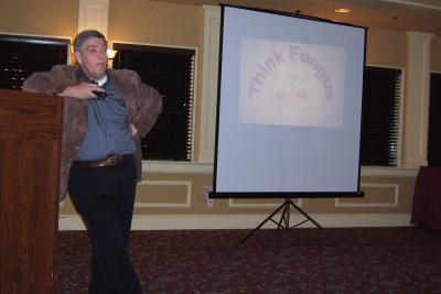
Michael Rinaldi, PhD. "Think Fungus"
back to top
Clinical Mycology Workshop
Bristol Community College, the Massachusetts Department of Public Health, the Northeast Branch, American Society for Microbiology and the National Laboratory Training Network co-sponsored a 2-day clinical mycology program on May 25 and 26th. The intermediate to advanced level program featured internationally renowned faculty from the Fungus Testing Laboratory at the University of Texas, Health Science Center in San Antonio. Instructors were Michael G. Rinaldi, PhD, Professor of Pathology, Medicine, Microbiology and Clinical Laboratory Science, Director of the Fungus Testing Laboratory; Annette W. Fothergill, MA, MBA, MT(ASCP), CLS(NCA), Assistant Professor, Technical Director, Fungus Testing Laboratory and Deanna Sutton, PhD, MT, SM(ASCP), RM, SM(NRM), Assistant Professor and Technical Director of Fungus Testing Laboratory.
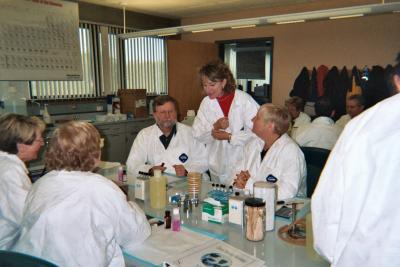
Annette Fothergill (C ) and Students
The combined lectures, demonstrations, case studies and laboratory exercises provided a comprehensive overview of current principles and practices in clinical mycology. Over forty-two clinical mycologists representing most major medical centers in Massachusetts, as well as professionals from Maine, New Hampshire, Rhode Island, Florida and Iowa participated in the program.
Preceding the program, on Tuesday, May 24th Dr. Rinaldi spoke at a NEB-ASM Dinner-Meeting at Vinny Testa's Restaurant in Dedham, Massachusetts. Financial support for this program was provided by Biorad and Hardy Diagnostics.
back to top
Nanogen Technologies: Applications for Infectious Disease and Genetic Testing
Nanogen Technologies: Applications for Infectious Disease and Genetic Testing was presented by Jeffrey Hawkins on March 29, 2005 at Vinny Testa's, Dedham, MA. Mr. Hawkins began by informing an audience of seventy-five people that as of June 2005, Nanogen will supplement their microarray product line, widely used in genotyping, by introducing real-time PCR (RTPCR). He listed some of the advances Nanogen has made in the area of infectious disease.
Mr. Hawkins explained the operation of a microarray by describing its use in the analysis for single-nucloeotide polymorphism (SNP). The microchip test site in the NanoChip� Electronic Microarray is 80�m in diameter, and is coated with a permeation layer of polyacrilamide gel containing streptavidin. Test sites on the array are electronically given a positive charge. PCR-amplified, negatively charged biotinylated DNA (amplicon) is attracted to those sites, passes through the permeation layer and embeds itself. Biotin in the amplicon DNA binds to the streptavidin in the gel, and the sample remains anchored at the test site after the electronic charge has been discontinued. Two fluorescent-labeled highly specific reporter probes are then hybridized to the DNA. The instrument scans the chip and from an analysis of the red and green fluorescent ratios can identify the SNP. If there is no initial match of probe with amplicon, increased stringency can be applied to re-testing. The sample can be washed off and another applied and tested. A blank array cartridge chip can be used in multiple ways; it can be used to test for multiple SNP's in a single sample, or to test for a single SNP in multiple samples. Both 100 and 400 site cartridges are available and multiple samples can be tested on each microarray without cross contamination, due to the electronic addressing system used in sample application. "Blank" cartridges are available for user-customized arrays. PCR-based research applications are currently available for the large NanoChip� Molecular Biology Workstation that is designed for research settings. Instrumentation and reagents for all tests are available from Nanogen. Mr. Hawkins passed around a microchip for the audience to view.
Testing for infectious diseases can be done using microarrays and Nanogen has developed specific probes to herpes simplex viruses 1 and 2, N. meningitidis, S. pneumoniae, and H. influenzae. Amplicons in these tests are permanently attached to probes, but can be detached and assayed for specific organisms. The Nanochip� 400 System has specifically been specifically designed for this application with the clinician in mind. It possesses a small footprint (microwave oven size), is fully automated, has a high throughput, and user-friendly software. Nanogen is also developing analyte specific reagents (ASR's) with which laboratories can develop tests for respiratory viruses. A single array will detect the sequences of and differentiate between six of the common respiratory viruses: influenza A, influenza B, parainfluenza 1, 2 and 3, and respiratory synctial virus A/B; additional assays are being developed. The tests will utilize the NanoChip� 400 System, with a single platform, multiplex amplification and detection protocols, biotinylated capture probes, fluorescence detection, and will be able to detect dual infections. Results will be available in one-day vs delayed culture-based tests.
Nanogen acquired Minor Groove Binder Technology (MGB) when it purchased Epoch Biosciences. The MGB technology will serve as Nanogen's entry into the area of RT-PCR. This technology, employing "superbases" that stabilize DNA probe-target interactions, enable the used of shorter very specific probes. Such probes provide the ability to distinguish between genetic variants of such rapidly mutating viruses as Influenza A and B. Mr. Hawkins showed and explained a diagram of the system in which a 400-site array is used. In the future, said Mr. Hawkins, we may see a decentralized, portable instrument, providing information from the time the sample is received to when an answer is obtained, thus enabling physicians to treat patients quickly and appropriately. In other areas, microarrays can be used for rapid identification of biological warfare agents, infectious disease organisms in water and food, and veterinary pathogens.
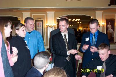
Jeffrey Hawkins and Norwich University
Students Examine a Microchip
back to top
Ticks & Tickborne Diseases in the Clinical Laboratory
The Northeast Branch, Massachusetts Department of Public Health and the National Laboratory Training Network co-sponsored a half-day program entitled Ticks & Tickborne Diseases in the Clinical Laboratory on March 15, 2005. The intermediate level program was held at MassBay Community College, Wellesley, MA and presented an overview of tick-borne diseases encountered in the Northeast and the epidemiology of tick-borne diseases in Massachusetts. Laboratory tests and algorithms used for diagnosis and patient management were discussed, and the laboratory session focused on appropriate methods for processing and identifying ticks. Faculty were Alfred DeMaria, Jr., MD, Assistant Commissioner and State Epidemiologist, Director, Bureau of Communicable Disease Control, State Laboratory Institute, MA Department of Public Health, Boston, MA; Richard Pollack, PhD, Laboratory of Public Health Entomology, Department of Immunology and Infectious Diseases, Harvard School of Public Health, Boston, MA; and Sam Telford, ScD., Associate Professor, Tufts School of Veterinary Medicine, North Grafton, MA.
back to top
40th Annual Regional Meeting
The 40th Annual Region I Branch Meeting of the American Society for Microbiology was hosted by the Connecticut Valley Branch on November 1-2, 2005. MEETING THE CHALLENGES was held at the Crowne-Plaza Hartford-Cromwell Hotel and Conference Center Cromwell, Connecticut. The Northeast Branch presented a half-day program which included the following topics: Group B Streptococcus: Physiology, Virulence and Vaccines, was presented by Dr. Lawrence Paoletti, Harvard Medical School, Boston MA; The Impact of Chromogenic Media on the Clinical Microbiology Laboratory by Dr. Michael Nugent, Becton-Dickinson (program sponsored by Becton Dickinson), and Rapid Analysis of Influenza A Viruses for Antiviral Drug Resistance Using Pyrosequencing was presented by Richard A. Bright, PhD, Influenza Branch, Centers for Disease Control, Atlanta, GA (program sponsored by Biotage).
back to top
57th Annual CLS/CNE Meeting
The 57th Annual Meeting of the Clinical Laboratory Science Society of New England (CLS/CNE) was held at the Rhode Island Convention Center in Providence on May 10-12, 2005. The NEB-ASM is pleased to have jointly sponsored the meeting along with four additional professional associations.
back to top
Career Night in Microbiology
Career Night in Microbiology was presented at the University of New Hampshire (UNH) on Thursday, April 7, 2005 and was designed to provide information about the types of positions available in this field. We would like to thank UNH faculty Prof. Thomas Pistole and Prof. Elise Sullivan for hosting this excellent student program for the second year in a row. Financial support was provided by the Northeast Branch-ASM.
Speakers and faculty hosts for UNH Career Night:
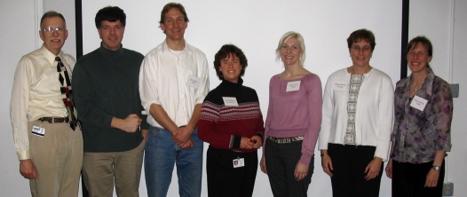
(L to R) Prof. Thomas Pistole, Dr. Patrick Regan,
Mr. Erik Janicki, Ms. April Blodgett, Ms. Kristen Weir,
Ms. Barbara Mullin, Prof. Elise Sullivan
Bethany Cooper, Manager of Employer Relations & Recruiting, University Advising and Career Center, opened the evening with information about the services available at UNH to students seeking employment.
Barbara Landau Mullin, B.A. in Microbiology from UNH and currently Worldwide Marketing Manager, Cell Culture Products, Corning Life Sciences, Acton, MA, spoke about her nearly thirty years of post-graduate experience, with a focus on marketing and sales.
Dr. Patrick Regan, Ph.D. in Microbiology from UNH and currently Supervisory Microbiologist at the Food and Drug Administration's Winchester Engineering and Analytical Center, spoke about job opportunities with the federal government.
Kristen Weir, B.A. in Biology from Kalamazoo College (MI), M.A. in journalism from New York University's Science and Environmental Reporting Program, and currently science writer for the New Hampshire Sea Grant Program at UNH, spoke about the joys of combining science and writing as a career.
April Blodgett, B.A. in Microbiology, UNH, and currently Senior Research Associate, AstraZeneca R & D, spoke about her experiences in both academic and industrial research.
Erik Janicki, B.A. in Biology, Colby College, former secondary school teacher, and currently M.S. candidate in Microbiology, UNH, spoke about his experiences in teaching at both the high school and college level.
During a break students chatted informally with the presenters and enjoyed pizza and soda.
back to top
The Future of Clinical Microbiology: Or Is It Clinical Chemistry?
Daniel Shapiro, MD, Director of Clinical Microbiology Laboratory at the Lahey Clinic, in Burlington, MA spoke on The Future of Clinical Microbiology: Or is it Clinical Chemistry? on May 5, 2005. An audience of eighty-seven microbiologists and other scientists attended the meeting at Vinny Testa's in Dedham, MA. He addressed the current and future role of molecular testing. Will such testing be a part of chemistry or microbiology? This is real question to microbiologists, as microbiological tests are rapidly being replaced by molecular biology.
Dr. Shapiro began with an overview of the history of bacteriology identification methods.
The clinical microbiology laboratory differs dramatically from clinical chemistry and hematology laboratories, he said. Microbiologists are dealing with living organisms with specific growth requirements. Identification frequently requires taking a probabilistic approach, where speed and accuracy tradeoffs are more obvious than with chemical tests. For any given hospital patient, chemistry and hematology laboratory work is completed when it is reported to physicians. However, for cultured sites, many procedures are done on a single original specimen.
From 1900-1945, identification was accomplished by a combination of physiological and biochemical tests, such as fermentation. Taxonomy and identification were not yet standardized. Gram negative organisms for example, were identified as Bacterium or Bacillus. Such "lumping" or "splitting" continue to be an issue in taxonomy to this day. By the mid 20th century, computers classified bacteria numerically using the results of biochemistry and morphological characteristics to assist in their identification using probabilities and probability charts. Dr. Shapiro then showed examples of such numerical methods and an identification matrix. An intuitive prior knowledge is required in order to be able to accurately interpret such probability concepts in conjunction with the likelihood of certain isolates occurring. Neither chemists nor hematologists have this experience.
Starting in the 1960's miniaturized versions of standard biochemical tests were introduced for bacterial identification (note: catalase, oxidase, etc. are chemical tests). However, phenotypic assays can result in incorrect results. Certain phenotypic features of bacteria may be dependent on environmental factors while tests in commercially available assays are fixed, even if new organisms are discovered. Therefore databases become less dependable and there is a tradeoff between timeliness and accuracy. There are numerous examples of such problems, such as F. tularensis being misidentified as Haemophilus and Actinobacillus, and Brucella melitensis being misidentified by API. Misidentification of Burkholderia cepacia can result in a downward course for cystic fibrosis patients, and Vitek yeast cards are only about 55% correct. You must read the literature to know such exceptions said Dr. Shapiro, since databases are not constantly updated.
Databases vary in the extent of their use of standard biochemical reactions, cell wall fatty acids, performed enzymes, and nucleic acid sequences for identification. Technology evolved and in the 1970's DNA:DNA hybridization was developed that enabled the creation of phylogenetic trees. Dr. Shapiro questioned if this is really a better way to find relatedness.
In the 1990's we had sequencing of 16S Ribosomal RNA (rRNA) genes that mutate at a slow, relatively constant rate. The various bacterial sequences are complied in databases. Dendograms are produced from the rRNA gene sequence enabling one to determine the relatedness of one organism to another in the database. Concerns and problems with these dendograms is that there are no guidelines as to what constitutes a new species on the basis of 16S rRNA sequencing. Sequencing errors occur and there is much intra-species variation. The 16S gene is only a small part of the genome even if conserved; other genes must be used in phylogenetic analysis. Pitfalls can occur using molecular methods for identification, for example, when a nucleic acid sequence that is used as a primer or probe is not unique. In the mycobacteria, M. celatum, type 1 differs in one base from M. tuberculosis complex. Temperature had to be changed to differentiate between the two. Dr. Shapiro again stressed the need to have a microbiology background and experience to initially detect such a problem in the microbiology laboratory.
Many times physicians request multiple tests on a single specimen. Multiple samples or multiple requests on a single specimen can be tested using microarrays that can have 100-400 tests laid out on a geometric grid, with each site being controlled electronically through a computer. Such methods are extremely accurate. However, with small volumes, the organism may not be detected even following amplification.
The ability to use the same platform for different genes or organisms will exert pressure to have a centralized molecular genetics laboratory. The question now arising is who is qualified to perform such molecular diagnostics testing as infectious disease tests, inherited disease tests, identification (paternity) testing, oncology, etc.? The AACC certifies people in this field, but the ASM does not; this could present a future problem. Microbiologists need to realize this, for soon regulatory/certifying agencies will require that only certified people can perform these tests.
The gold standards (reference methods) for susceptibility testing are either the Kirby Bauer disc method or antibiotic dilutions (MICs). Problems arise when a reference method indicates a microorganism is susceptible to a drug and the new method being evaluated indicates it is resistant. The laboratory must try to minimize errors as much as possible. Since there are many different and novel mechanisms of resistance worldwide, we will continue to use phylogenetic testing for identification using specialized media and susceptibility testing.
It appears that molecular methods will be the choice for identifying pathogens in the future said Dr. Shapiro. However, the speed with which new mechanisms of drug resistance are evolving will see to it that no single institution will have a complete library of organisms, and libraries become outdated rapidly. But will these be the methods of choice (even if they are cost effective) for the identification of bacteria, viruses and fungi?
Therefore asked Dr. Shapiro, what is the future of microbiology and chemistry? In his opinion, molecular diagnostic testing will have its own laboratory area and microbiology will be only one part of it. Classical virology (tissue cultures, shell vial assays, etc.) will be non-existent. The probable bacteriology laboratory workflow will be from isolation to molecular laboratory to susceptibility tests for Gram negative organisms. A number of phenotypic tests will be performed, and the results will be confirmed by genetic tests. However, who will certify molecular identification tests? And who will actually perform molecular identification? Will they need to be medical technologists? Or personnel with a Bachelors degree in biology, who had laboratory-based classes in which molecular methods were used? The certification of laboratory directors is also unclear. The future of microbiologists is still up in the air!
back to top
Food Microbiology: Moving Into the Molecular Testing Era
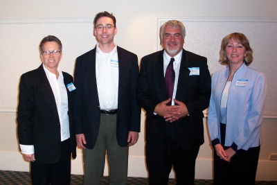
Patricia Rule, Peter Carlson, NEB President
Gregory Reppucci and Nellie Dumas
A half-day symposium, Food Microbiology: Moving Into the Molecular Testing Era was held on April 14, 2005 at the Radisson Airport Hotel, a Johnson & Wales University Education Facility, in Warwick, RI. Fifty-three people attended.
Patricia Rule, Senior Staff Scientist at bioMerieux, Inc. started the symposium with VIDAS, Next Day Solutions for Rapid Food Pathogen Detection. She joined the bioMerieux Research and Development team in 1987, contri-buting to test kit development for the Vitek�, Vidas� and Bactometer� used in the food and pharmaceutical industries. Clinical and food microbiology products marketed by bioMerieux involve partnerships with Affymetrix, Gen-Probe, MIDI, Organon Teknika and alliances with other groups. Products marketed by bioMerieux include prepared media, Vitek�, Vidas�, airIDEAL, api�, BacT/Alert, and Bactometer�. The products and instruments are used in industrial applications, research, blood and tissue banks, food and beverages industries, in pharmaceutical products, and cosmetics.
Foodborne illness causes 1000-3000 deaths/year, with 6.8-8.1 million cases/year, with an enormous financial cost. Ms Rule explained how Vidas � works in the rapid detection of food pathogens. This fully automated immunoassay system uses standardized enzyme-linked fluorescent assay (ELFA) technology, and can perform single or batch testing, with serology and immunochemistry results being available in 20-60 minutes. Each Vidas� assay consists of a strip that contains all reagents used for the reaction and a Solid Phase Receptacle, a plastic pipette-like device whose inside is coated with polyclonal and monoclonal antibodies. There are three steps in rapid pathogen screening (1) immunoassay, in which antibody captures target pathogens, (2) the Sandwich Test in which two antibodies are conjugated with enzymes and (3) color (florescence) development.
Ms Rule explained the process a manufacturer goes through in order to obtain AOAC certification for new instruments and reagents. The Vidas� LSX, LMO2, ICS and ECO assays were validated and certified by the AOAC Research Institute as being effective methods for the detection and selective isolation of Listeria species, Listeria monocytogenes, Salmonella species and E. coli 0157.
The Vidas� Immunoconcentration Salmonella (ICS) assay can detect Salmonella in all foods. It uses ICS broth with a selective plate method (Hektoen Enteric, Bismuth Sulfite, Salmonella Identification)-SMID and gives a Next Day solution. A 48 hour solution is available using the Vidas� Salmonella (SLM) assay that employs Tetrathionate and Selenite Cysteine broths.
The Vidas� E. coli (ECO) assay can be used for the automatic detection of E. coli 0157:H7. It uses automatic immunoconcentration and automatic 0157:H7 chromogenic agar with a same-day (8 hour) and next day (24 hour) solutions. On chromogenic medium E. coli is a specific blue-green color due to enzymatic activities. Other assays available for food pathogens are a 2-day assay for Campylobacter, and a same day assay for Staphylococcal enterotoxin II.
The Vidas� Listeria Species Xpress (LSX) with Ottaviani Agosti Agar (OAA) provides Next Day detection and selective isolation of Listeria in meat, dairy samples and on environmental surfaces. LX broth is used for optimized enrichment. One and two-step methods are used for Listeria isolation, but do not use cold enrichment, which takes too long. Listeria is pH, salt and cold tolerant, and after enrichment the pH in the enriched broth drops and Salmonella die while Listeria survives. OAA chromogenic agar is used to confirm results obtained with the Vidas � tests. Listeria species are turquoise on the agar, while L. monocytogenes or novii are turquoise with a halo behind them. Results for meat and dairy products are available in 30-34 hours after sampling, while environmental results are available in 26-28 hours. External laboratory studies have shown that it is hard to capture the organism all the time; detection becomes harder as the number of organisms gets lower (mpn/g: 0.023, 0.0036).
The identification of an organism isolated by Vidas� is confirmed by Vitek� or api�. The Vitek automates all steps needed to identify gram positive and negative organisms. A miniature card format (Vitek card) consisting of 32-480 wells (depending on the model) and fluorescent technology are used to identify the organisms. The system use state-of-the-art technology and provides rapid and accurate identification of food pathogens and can also be used for antimicrobial susceptibility testing.
Peter Carlson, Senior Sales Representative at Cepheid, spoke next on Detection and Quantification of Foodborne Pathogens Using Real-Time PCR. Cepheid develops, manufactures and markets fully integrated systems that perform genetic analysis for the clinical, life sciences and biothreat markets. Cepheid is a young company, whose sole focus is real time-PCR (RTPCR). The Company was involved in developing platforms for anthrax detection for the postal service after 9/11.He listed the most frequently isolated food pathogens as E. coli, Salmonella, Vibrio, Norwalk-like viruses, rotavirus, hepatitis A, and Cryposporidium parvum. The BAM methods in general use for microbiological testing of food products are characterized as being slow and involve selective growth on media. When coupled with sensitive and specific ELISA tests, identification can be completed in 24-48 hours. Classical PCR, on the other hand, provides for specific same day identification. Real time-PCR (RTPCR) is more rapid than classical PCR and provides for even greater sensitivity and specificity. RTPCR permits direct quantification testing to be carried out in less than 30 minutes. If prior extraction/concentration steps are used, the organisms can be can detected in less than 1 hour. In RTPCR, the target is amplified in the presence of a reporter, an instrument excites and detects the reporter, with signal intensity being directly proportional to the amount of amplified DNA. The threshold cycle (CT) is a fractional cycle at which target-signal reaches the threshold. RTPCR is better for quantitation than conventional PCR because it looks at the baseline and exponential parts of the growth curve while conventional PCR looks at the linear log (plateau). Serial dilution can be used to create a linear standard curve to determine the concentration of organisms in a sample.
Detection chemistries that can be used with the SmartCycler include sybr green I and ethidium bromide which bind to double-stranded DNA. Detection involves labeled probes/ primers, molecular beacons, and fret probes. Cepheid's Smart Cycler has up to 96 independently programmable reaction sites, making it highly flexible. The I-Core � Module, an independently-controlled thermal cycler fluorometer, permits multiplex PCR as a result of its four-channel optical detection. Optical blocks are designed to simultaneously excite and detect four different spectral bands.
The preparation of food samples for analysis is a critical step said Mr. Carlson, since various inhibitors may be present. Inhibitors become more of a problem when a sample must be concentrated due to the presence of low number of organisms. Using the sample directly, with no extraction, permits the quantification of organisms. Cepheid is currently focusing it efforts on developing a method for Vibrio, which causes one 1 of 160 food related deaths in the US, with Vibrio vulnificus causing most of the deaths. Vibrio parahemolyticus outbreaks are also increasing; this organism causes gastroenteritis lasting 2-5 days and is found in wound infections.
BAM Vibrio detection uses homogeneous enrichment that selectively isolates the organisms, and takes days. Multiplex PCR on the other hand, targets the specific genes tl, tdh, trh. It can be used for food and environmental cases. Specific assays should be able to detect down to 1 cfu/ml and can be run in an easy package. Detection can be completed in less than an hour using 4-channel multiplex amplification. In summary, said Mr. Carlson, RTPCR can be a valuable tool for the rapid identification of foodborne bacteria in water and food.
Ms Nellie Dumas, Supervisor, Clinical Bacteriology Laboratories of Wadsworth Center, New York State Dept. of Health concluded the symposium with Rapid Identification in the Public Health Investigation of Foodborne Outbreaks in New York State. She explained why rapid testing is important as she reviewed the roles of the NYS Laboratories in the investigation of foodborne outbreaks.
The NY Disease Surveillance System functions as an immediate response system, as well as to watch long term trends, to identify new problems and guide administrative priorities and policies. Many people are involved in the actual investigations, with epidemiologists and laboratorians (hospital, commercial, city, county, state and federal) at the center. Also involved are city and county health departments, state agencies and such federal agencies as the Centers for Disease Control and Food and Drug Administration.
Each state has its own reportable disease laws, mandating that physicians, hospitals, schools, etc. must report communicable disease incidents to the local health departments. In NY, positive results are confirmed and linked to cases at Wadsworth by pulse field gel electrophoresis (PFGE) and placed on the state information network. Local health departments do the investigations and report their findings to the state. They also send case reports to the state. Food outbreaks can occur as single events (wedding, reception, banquet, church supper), or clusters (time/space clustering of laboratory-confirmed cases) for which PFGE is valuable to link cases, or ongoing incidents (related to a food service establishment or contaminated products) which are detected by several complaints to local health departments or to a communicable disease section. The number of E. coli and Listeria cases in NY has decreased since 1980, but Salmonella have not. The etiology of most foodborne outbreaks remains unknown, but E. coli and Listeria are prime suspects, viruses are second and Salmonella are third.
Steps that epidemiologists take in an investigation include surveillance, i.e. identifying the outbreak, outbreak investigation, and outbreak control, followed by prevention and education. Also, many epidemiological questions are incorporated into laboratory analysis forms (person, place, time, epidemiological data), so that a hypothesis regarding the outbreak can be generated. All agencies work together on an outbreak.
Ms Dumas then told of a foodborne outbreak involving cider at a specific orchard. She defined a case as anyone with symptoms, regardless of their county or state of residence; this definition crosses state lines. The main symptoms were diarrhea, bloody diarrhea, abdominal cramps, in 14 hours, people ages 1-55+ (most were young). Forty-three people visited an orchard on a weekend and started to get ill on Monday of the next week. The county of residence and date that each case visited the orchard were noted. However, some people purchased the cider to take home to drink. In all there were 46 controls and 23 cases. Controls were defined as people who were at the orchard in September, had consumed food, but were not ill.
Conference calls were set up between all agencies involved. Hospitals looked for E. coli 0157:H7 but found none. More conference calls ensued, and by September 29th, the cider press was close, some agencies wanted to close entire orchard. Conference calls as to what to do continued, as did the investigation.
Bacteriological tests used for identification of organisms included microscopic and macroscopic tests, conventional biochemicals, serology, an immunomagnetic separation system, and DNA sequencing. Specimens tested were patient isolates, stools, environmental swabs and swipes, the apple hopper and cider press. Fecal specimens were taken from calves and horses. Both opened and unopened apple cider from the orchard and from patient homes were tested. People working the cider press also took care of the animals and two refused to give samples. Contamination seemed to come from animals to people to the cider.
Real-time PCR for Shiga toxin 1 and 2 was performed, also PFGE. The organism was sorbitol positive, not negative. Now the question was whether the problem was caused by another serotype. Immunochromatography (ELISA), immunomagnetic bead separation (SMAC) and CMAS medium were used, but toxigenic E. coli could not be isolated/identified from the cider. How was the source confirmed? DNA fingerprinting by PFGE showed a number of serotypes of 011 in calves, which showed about ten different PFGE patterns, none of which were recovered from the patients.
However, Cryptosporidium produces symptoms similar to that of E. coli. The infective dose is only 10-100 oocysts. The same samples that were tested for E. coli were also tested for Cryptosporidium using concentrated acid-fast smears; Cryptosporidium was thus confirmed in the cider, where fermentation doesn't affect it.
Of great value in foodborne outbreaks is the Pulse Net (CDC) National Molecular Subtyping Network for Foodborne Disease surveillance, where information is electronically posted so that states can compare PFGE results. Ms Dumas again emphasized that communication is a major part of laboratory work, and it takes lots of teamwork to solve an outbreak.
back to top

|
|
|